Development of an Image Diagnosis Technology Utilizing a High-Sensitivity Magnetic Sensor
Yokohama National University, a national university corporation (Yokohama-city, Kanagawa pref. President: Izuru Umehara, hereinafter “YNU”) and TDK Corporation (Tokyo, President: Shigenao Ishiguro, hereinafter “TDK”) have developed a prototype image diagnosis technology utilizing a high-sensitivity magnetic sensor*.
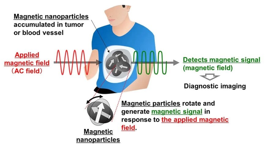
The developed prototype technology is related to the magnetic particle imaging* method which is intended to detect and create images of magnetic particles* accumulated in a tumor or blood vessel.
Magnetic resonance imaging (MRI) diagnostics* and X-ray computerized tomography (CT) scanning* are used in clinical services in the diagnosis of organ health, tumors and other conditions using the contrasting density of imaged objects. In contrast, the magnetic particle imaging is intended for use in detecting only the tracers* in the imaged objects to create images similar to positron-emission tomography (PET)* and other similar technologies.
The principle of the magnetic particle imaging is to detect the magnetic signals generated by magnetic particles accumulated in a tumor or blood vessel from outside of the body (Figure 1). When intended for use in medical imaging, it is important for devices to be highly sensitive to enable the detection of small amounts of magnetic particles. Though magnetic particle imaging technologies primarily use a method that measures electromotive force electromagnetically induced through detection coils, the new technology developed by YNU utilizes a prototype high-sensitivity magnetic sensor to achieve this. The prototype high-sensitivity magnetic sensor was developed by TDK for use in the detection weak magnetic fields at room temperature.
Although still under development, the prototype sensor has been shown in a prior feasibility study to measure magnetic field distribution in a heart.1 Through this development, the sensor has successfully reduced the strength of the alternate current magnetic fields applied from outside of the body to one tenth lower than conventional levels. This reduced strength of the applied field is achieved by the non-linear response characteristics of the sensor to the measured magnetic field strength.
From this achievement, it is expected that the utilization of high-sensitivity magnetic sensors will enable magnetic particles to be detected across wider imaging ranges including the head or whole body of a human.
Going forward, YNU and TDK will continue to develop this technology, with the goal of creating magnetic particle imaging devices that can be used practically in clinical services.
1 2019 feasibility study conducted using a prototype 99-channel sensor array in five healthy human subjects.
Glossary
- High-sensitivity magnetic sensor
A magnetoresistance effect-based magnetic sensor (MR Magnetic sensor) developed by TDK that uses the Nivio xMR sensor. It has a compact sensor head and can also detect biosignals at room temperature. Its magnetic field detection performance almost reaches that of the superconducting quantum interference device (SQUID) flux meter, which requires cooling.
- Magnetic particle
Also called magnetic nanoparticles, they are expected to be applied in magnetic particle imaging and hyperthermia cancer therapy. Typically, these particles are iron oxide(Fe3O4 and γ-Fe2O3) about 10 nm in diameter that are used in practice as contrast agents for MRI because of their biological compatibility.
- Magnetic particle imaging
A new diagnostic imaging method proposed in 2005. It detects and creates images of magnetic particles accumulated in a tumor or blood vessel from outside of the body (Figure 1). The devices for small animals (animal testing) are available commercially from Europe and the U.S. However, a device for clinical use on humans has not been developed yet.
- Tracer
Materials that are injected in the objects to be imaged for detection, to observe organs or tumors. These can be radioactive isotopes used for PET and magnetic particles for magnetic particle imaging.
- (Related information) Magnetic resonance imaging (MRI) diagnostics, X-ray computerized tomography (CT) scanning
MRIs detect and creates images of hydrogen atoms in organs while X-ray CT creates tomographic images from X-ray images taken from multiple directions. Both technologies are used for the diagnosis of organ health, tumors and other medical conditions based on contrasting densities in an image.
- (Related information) Positron-emission tomography (PET)
It combines radioactive isotopes of materials such as glucose and detects the radioactive isotopes from outside the body to create diagnostic images based on the distribution of the radioactive glucose or other tracer in the body.
www.tdk.com